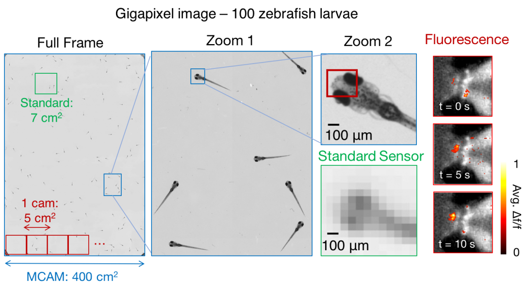
Capturing video with gigapixel-sized frames using nearly 100 lenses and sensors
We have been developing a suite of multi-camera array microscopes for a variety of biomedical imaging applications. More information about this new technology may be found at https://www.ramonaoptics.com/
We also have an associated project page that we will keep updated with interesting data and videos: mcam.deepimaging.io
Please check out out gigapixel video viewer as well!
Associated papers:
X. Yang et al., "Curvature-adaptive gigapixel microscopy at submicron resolution and centimeter scale," Optics Letters (2025).
A. Chaware et al., "Micron scale 3D imaging with a multi-camera array," Advanced Imaging (2025).
C. Cook et al., "Fourier lightfield multiview stereoscope for large field-of-view 3D imaging in microsurgical settings," Advanced Photonics Nexus (2025).
L. Kreiss et al., "Recording dynamic facial micro-expressions with a multi-focus camera array," Biomedical Optics Express (2024).
K. C. Zhou et al., "High-speed 4D fluorescence light field tomography of whole freely moving organisms," Optica (2025).
K. Kim et al., "Rapid 3D imaging at cellular resolution for digital cytopathology with a multi-camera array scanner (MCAS)," NPJ Imaging (2024).
K. Zhou et al., "Parallelized computational 3D video microscopy of freely moving organisms at multiple gigapixels per second," Nature Photonics (2023). Pre-print available here. Project page available here.
M. Harfouche et al., "Imaging across multiple spatial scales with the multi-camera array microscope", Optica (2023). Pre-print available here. Project page available here.
X. Yang et al., "Multi-modal imaging using a cascaded microscope design", Optics Letters (2023). Pre-print available here. Project page available here.
E. Thomson et al., "Gigapixel imaging with a multi-camera array microscope," eLife (2022). Project page available here.
Project Summary:
High-throughput optical microscopy is currently transforming the research fields of genetics, drug discovery and neuroscience. Due to challenges with large lens design, standard microscopes have a hard time capturing anything more than 50 megapixels per image snapshot, which makes it impossible to simultaneously image at cellular-resolution over a multi-centimeter viewing area (field of view, FOV). To enable cellular resolution imaging over fields of view 10s of cm in diameter, we are utilizing an array of miniaturized cameras, tiled in a 12x8 configuration and collectively called the micro-camera array microscope (MCAM).

Each camera is individually addressable and controllable to enable rapid acquisition over the fields of view as large as 20cm in diameter, providing us with gigapixel-scale snapshot images. Preliminary applications for the MCAM are being developed for behavioral imaging of freely swimming zebrafish larvae. This work is in collaboration with Ramona Optics, Prof. Eva Naumann in the Duke School of Medicine, Dr. Tim Dunn in Duke Statistical Science, and a number of other important collaborators!
Please check out some of our new gigapixel videos at our online interface here: gigazoom.rc.duke.edu

If you are interested in our extreme wide-field-of-view microscope and think you have an interesting application for us, please feel free to get in touch!